Save up to 60% on your scans and tests bookings. Book now

Scan type
Sound wave-based

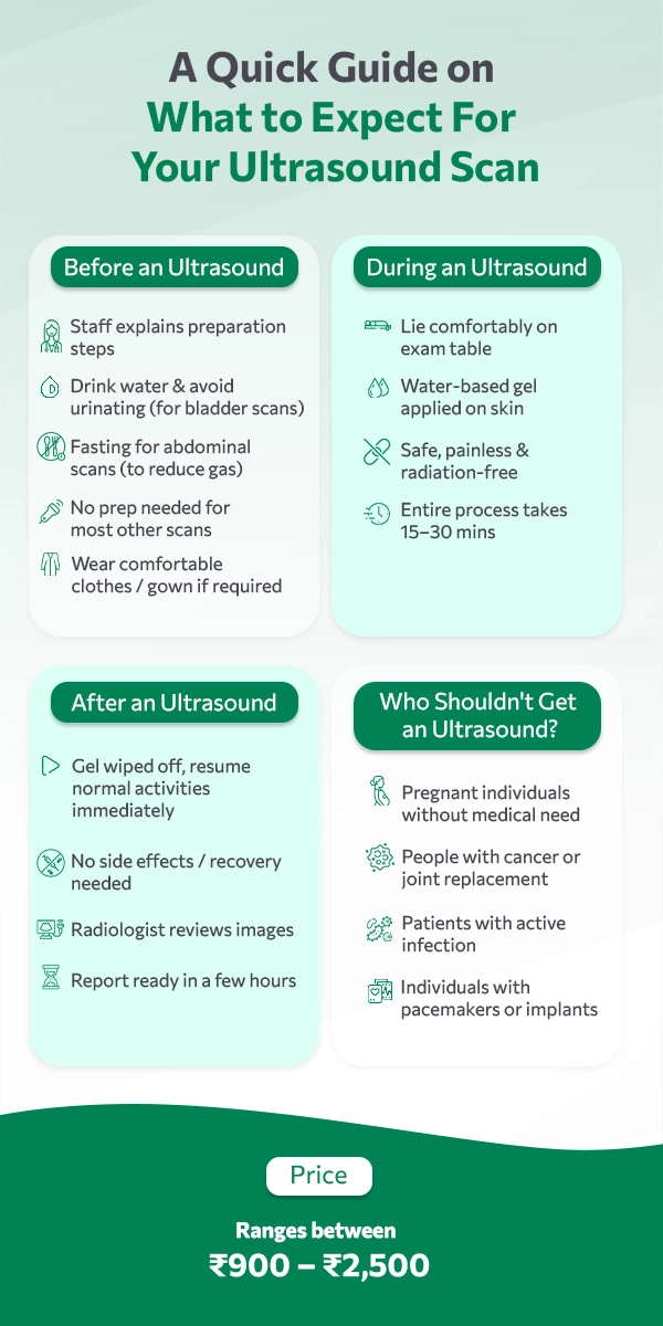
Duration
15–30 minutes
Report TAT
6 hours

Price
₹1,300 – ₹3,900
Radiation risk
No radiation

Serving a large patient base allows us to keep costs low and pass on the benefit directly to patients.

Automation and digital workflows reduce manual work in reporting, improving speed while cutting costs.

Our leadership operates with in-depth knowledge of local needs, ensuring effective and cost-conscious operations.

We invest wisely in facilities and real estate, avoiding unnecessary overheads.

Every rupee is spent with care, prioritizing patient benefit over extravagance.

We ensure that our savings is passed directly to the patients instead of intermediaries.

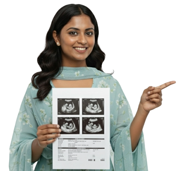
Detailed pregnancy scan to check baby’s growth and detect abnormalities.
View More
Clear imaging of female reproductive organs for fertility or pelvic issues.
View More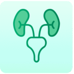

Early pregnancy scan to assess baby’s health and screen for chromosomal issues.
View More
Blood flow imaging to assess arteries, veins, and circulatory health.
View More
Focused pelvic scan for reproductive organs, bladder, and related conditions.
View More
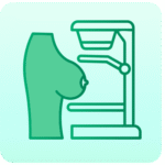
Safe abdominal scan to assess liver, pancreas, gall bladder, and more.
View More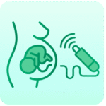
Routine pregnancy scan to monitor the baby’s development and the mother’s health.
View More
Essential pregnancy scan for fetal growth, position, and well-being.
View More
Pregnancy scan to measure the baby’s blood flow and placental health.
View More