Ever wondered about the inner workings of PET scans? PET Scan is one of the vital diagnostic tests. Understanding how PET scans operate is crucial for making informed decisions regarding your health. Join us to learn about the mechanics of PET scans!
Positron Emission Tomography (PET) is a nuclear medicine imaging method that uses radioactive tracers to observe organ function in real time. In this technique, a PET scanner emits positrons—particles interacting with tissues and generating gamma rays. PET scanner captures gamma rays, constructing images that show the cellular metabolic activity. It diagnoses various conditions such as cancer, heart disease, and brain disorders effectively. It also measures essential functions like blood flow and blood sugar metabolism.
They are particularly effective at showing areas of increased metabolic activity. PET scans reveal small tumors which might go undetectable on other diagnostic tests such as CT or MRI.
Preparations required for PET Scan
- Typically, you’ll need to fast for several hours (usually 6-8 hours) before the PET scan, especially if the scan is around abdominal area.
- Refrain from intense physical activities for at least 12 hours before the scan to ensure accurate results.
- Consult with your healthcare provider regarding any medications you are taking, as certain medications may interfere with the scan.
- Drink plenty of water to stay well-hydrated before the scan, unless instructed otherwise by your healthcare provider.
- Wear comfortable clothing without metal zippers or buttons, as you may be asked to change into a hospital gown.
- Notify if you are pregnant or breastfeeding, as special precautions may be necessary.
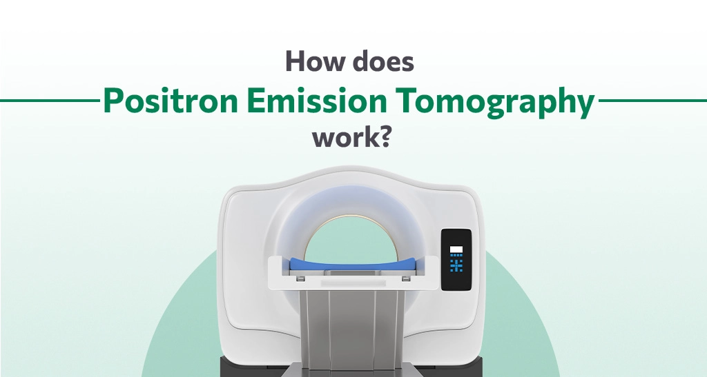
How does the scan work?
A PET scan is a type of nuclear medicine imaging method utilizing minimal, safe doses of radioactive substances known as radiotracers. It is administered through an IV and introduced into the body. This radioactive glucose variant is infused into a vein, and the PET scanner captures images. Once it is injected, the patient undergoes whole-body imaging with the PET scanner.
The purpose of a PET scan is to detect malignant cysts and tumors that was essentially missed or challenging to characterize using conventional CT scans, X-rays, or MRIs. Unlike traditional imaging methods, PET scans visualize biological processes and molecular activity within the body, providing the potential to identify diseases in their early stages.
Diseased cells in our bodies have higher absorption of the radiotracer compared to healthy cells, creating areas known as “hot spots.” The PET scanner detects and captures the radiation emitted from these hot spots, generating images of the affected tissue. For a more comprehensive examination, a PET/CT scan merges CT scan with the PET scan images.
What should you expect during the scan?
- Once you’re prepped, blood sugar levels are checked and then the radioactive tracer is injected into your vein through the intravenous line.
- You’ll be made to sit in a chair for about an hour while the radiotracer goes through the bloodstream and gets absorbed by your organs and tissues.
- You must remain still during the scan. However. any movement will blur the image.
- The scanner makes buzzing and clicking sounds as it takes the images.
PET scans are an important tool in the field of diagnosis to detect several diseases from cancer to heart diseases. They provide more precision and enhanced diagnosis because of their usage of radiotracers. So next time you’re going into a PET scan with knowledge about the procedure and working of PET scans, you can make more informed choices and ask the right questions.