Leave your name & phone number with us


CERTIFIED NABL LABS

200+ LABS ACROSS INDIA

1.5 CRORE PATIENTS SERVED
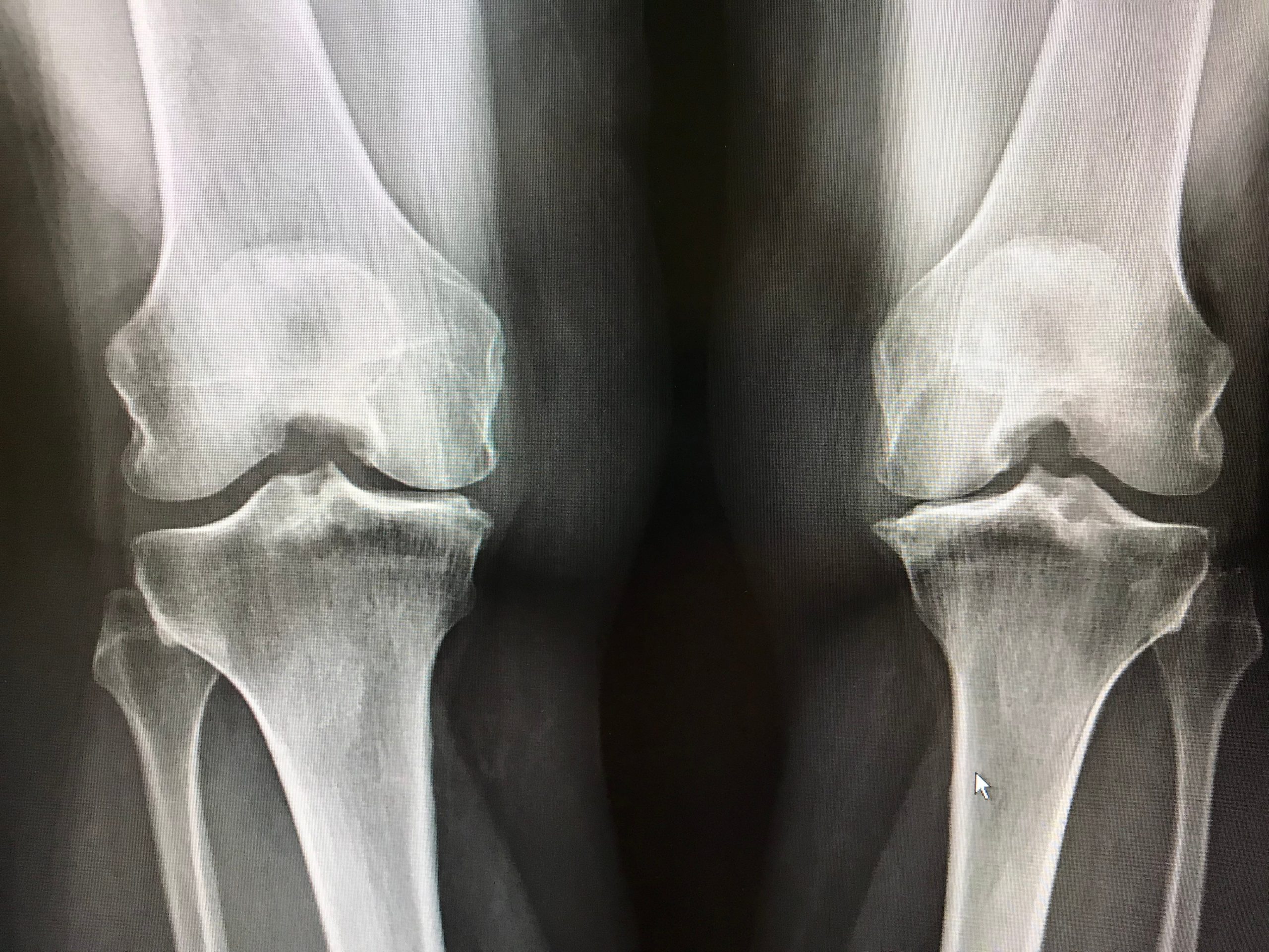
What is an Knee AP View X-ray?
A knee AP view x-ray is a type of x-ray that provides an anatomically accurate image of the knee joint and its surrounding soft tissue. This x-ray helps in diagnosing various musculoskeletal conditions such as fractures, dislocations, ligament tears, and infections. The x-ray is taken in the standing position with feet parallel to each other and knees bent at a right angle. By comparing both sides of the x-ray, medical professionals are better able to determine any abnormalities present to cause different orthopaedic ailments. A knee AP view x-ray is indispensable in properly guiding treatment plans as it gives a clear image of how the bone elements interact with each other at different angles, making it easier for doctors to accurately diagnose and treat patients efficiently.
When is a Knee AP View prescribed?
The Knee AP View x-ray is prescribed to review the knee joint for diagnosis and treatment of medical conditions. A knee x-ray can provide many insights, from tracking the progressions or regressions of chronic ailments, to diagnosing acute diseases or issues. The x-ray views may also be performed before or after a surgical procedure. An x-ray is one imaging tool available to assist in properly assessing an orthopaedic condition causing pain or discomfort in the knee joint. Generally speaking, it is best to consult a health care professional to get proper guidance regarding x-rays for diagnostics and treatments plans.