Leave your name & phone number with us

CERTIFIED NABL LABS

200+ LABS ACROSS INDIA

1.5 CRORE PATIENTS SERVED
What is an Pelvis MRI Scan?
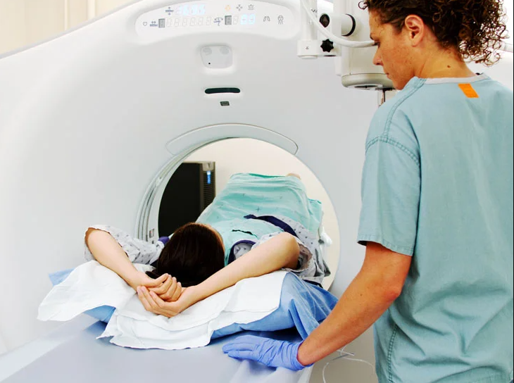
To get a clear picture of the pelvis, you may need to undergo an MRI scan. This imaging technique uses magnetic fields and radio waves to produce detailed images of the interior structures of the pelvis (uterus, ovaries, prostate, rectum and anal canal) , including those that make up the pelvis. This data can then be combined with other medical images to produce a complete picture of your pelvic anatomy, providing diagnostic information regarding any injuries or conditions that might be present in this area. If you are experiencing symptoms related to your pelvic region, such as pain or incontinence, it is important to speak with your doctor about undergoing an MRI scan for the pelvis in order to better understand what may be causing these symptoms and how best to treat them.
When and Why MRI Scan for Pelvis is Prescribed?
When it comes to assessing the health of the pelvic area, there are a number of different imaging techniques that can be used. In general, however, an MRI scan for the pelvis is most commonly recommended in circumstances where more specific information about a patient’s condition is required like uterine fibroids, ovarian cysts, prostate issues . Some common indications for an MRI scan include the presence of pain or discomfort, significant changes in bowel or bladder function, or other red flags such as unexplained weight loss. These scans are also commonly used to help assess and monitor the progression of certain chronic conditions like inflammatory bowel disease or pelvic floor disorders. Ultimately, an MRI scan for the pelvis provides much-needed insights and information that can help to guide treatment planning and improve patient outcomes.


