Leave your name & phone number with us

CERTIFIED NABL LABS

200+ LABS ACROSS INDIA

1.5 CRORE PATIENTS SERVED
What is an Pelvis Ultrasound?
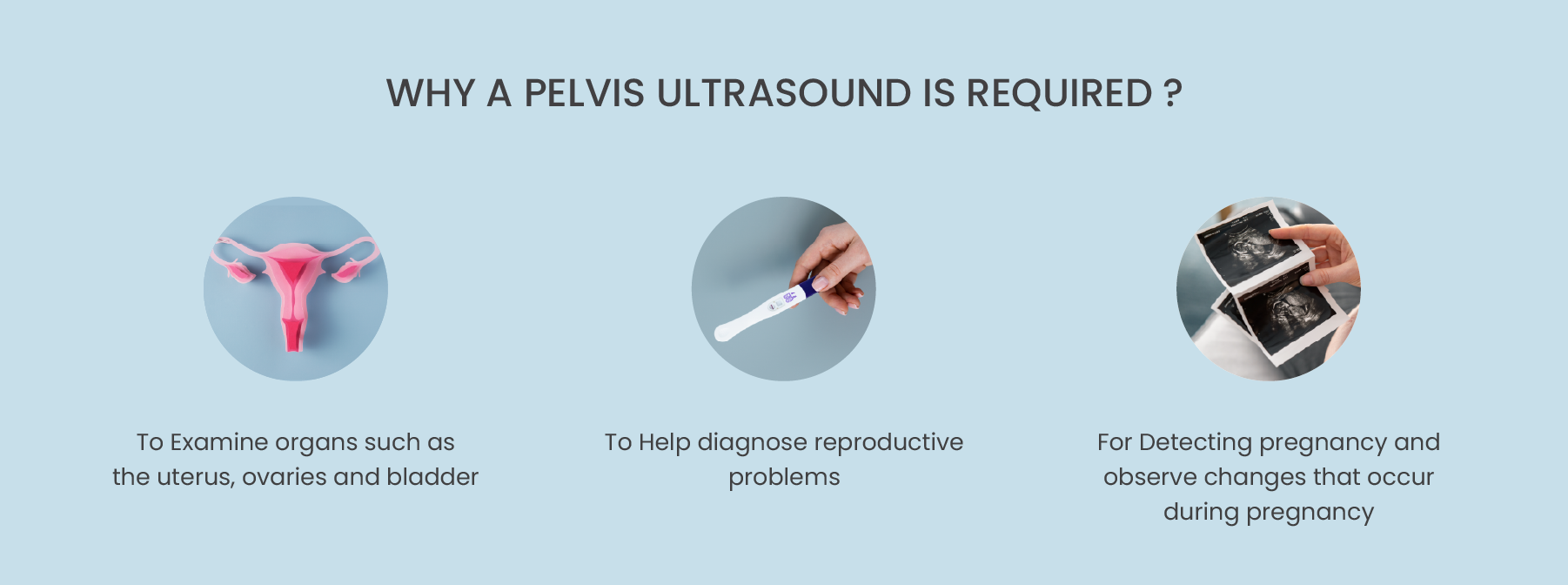
A pelvis ultrasound scan is a type of imaging technique that utilizes sound waves to create an image of the pelvic organs and structures. The pelvic area can be accessed via abdominal, vaginal or rectal ultrasound scanning depending on what information the physician requires. Pelvic ultrasound scanning is used to examine organs such as the uterus, ovaries and bladder in order to diagnose any potential problems these organs may have. It can also provide valuable information about neoplastic lesions, cysts, tumors or other abnormalities located in this area of the body. Pelvis ultrasound scans are simple examinations that provide immense insight into the health of the pelvic region with minimal discomfort and no radiation exposure.
When is a Pelvis Ultrasound prescribed?
Pelvis ultrasounds are becoming commonplace in the gynecological community, offering a non-invasive way for physicians to assess the internal organs of a woman’s pelvis. Pelvic ultrasounds can help diagnose reproductive problems, such as tumors or cysts in the ovaries or uterine fibroids, and detect abnormalities with ovarian or uterine physiology. Pelvic ultrasounds can also detect pregnancy and observe changes that occur during pregnancy, such as size or location of the fetus, and evaluate placenta positioning. Pelvis ultrasound scans are an invaluable diagnostic tool providing health practitioners with a comprehensive view of the uterus, ovaries, cervix, adnexa and/or bladder.


