MRI and CT are the two most widely used diagnostic tools; playing pivotal roles in assessing various organs, specializing in evaluating brain health. While distinct in their approaches, both of these techniques excel at accurately identifying abnormalities and neurological issues. Join us in this blog as we understand the intricacies of how MRI and CT contribute significantly to ensuring your neurological well-being.
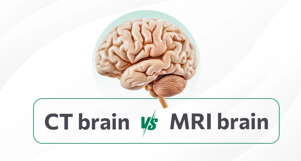
CT Brain
A CT scan surpasses standard X-rays as it offers more intricate cross-sectional images of brain tissues from various angles. Whether with or without a contrast agent, CT scans precisely assess brain function and are instrumental in identifying abnormal growths, tumors, lesions, or structural anomalies. Excels in highlighting bone structures and is efficient in detecting acute bleeding or fractures. However, it may offer less detailed images of soft tissues. Moreover, Brain CT aids in guiding surgeries and biopsies, as well as monitoring treatments and detecting blood clots within the brain.
MRI Brain
MRI takes the lead in neurological diagnostics. From identifying skeletal metastases to soft tissue intricacies within the brain, making it superior for assessing the brain’s anatomy and detecting abnormalities. It uses strong magnetic fields and radio waves to generate detailed images. Unlike CT scans, MRI brain scans do not use ionizing radiation. Its primary objective is to identify a spectrum of abnormalities, from masses and hemorrhages to inflammation and structural problems. Therefore, brain MRI plays a crucial role in cases where neurological conditions pose diagnostic and treatment challenges.
Navigating scan procedures
The Brain MRI procedure follows the standard MRI process, but with an emphasis on the head. We place a specialized helmet-like device equipped with coils and wires for transmitting brain activity on the patient’s head. After changing them into a comfortable hospital gown, we begin the scan. The 20- to 30-minute duration requires remaining perfectly still for clear and accurate images.
After changing into a hospital gown, IV injection is administered for contrast. Lying on a table, the head enters the machine, and as it rotates, X-ray beam images are captured and transformed into 3D pictures. The entire CT scan takes around 5–10 minutes to complete, making it both effective and efficient.
In conclusion, both CT Brain and MRI Brain serve as essential diagnostic tools, each with its own unique strengths. The choice between them depends on the specific requirements of the patient’s condition. With CT excelling in certain scenarios and MRI providing superior imaging for detailed assessments of brain anatomy and soft tissues. The treating physician guides the decision. He/she ensures that the chosen method aligns with the patient’s needs for accurate diagnosis and treatment planning. Schedule your appointment with Aarthi Scans and Labs.