fbpx
CERTIFIED NABL LABS
200+ LABS ACROSS INDIA
1.5 CRORE PATIENTS SERVED

ULTRASOUND SCAN IN ASL_BRANCHESAYAPPAKKAMARRAYPAGE2

An Ultrasound scan (or) USG can image the anatomy and pathology of organs in our abdomen, and neck. Ultrasound is also used to image for anomalies during pregnancy and to monitor the growth of the baby. Colour doppler scans are used to image the flow within arteries and veins in our body.

Ultrasound Scan Procedure

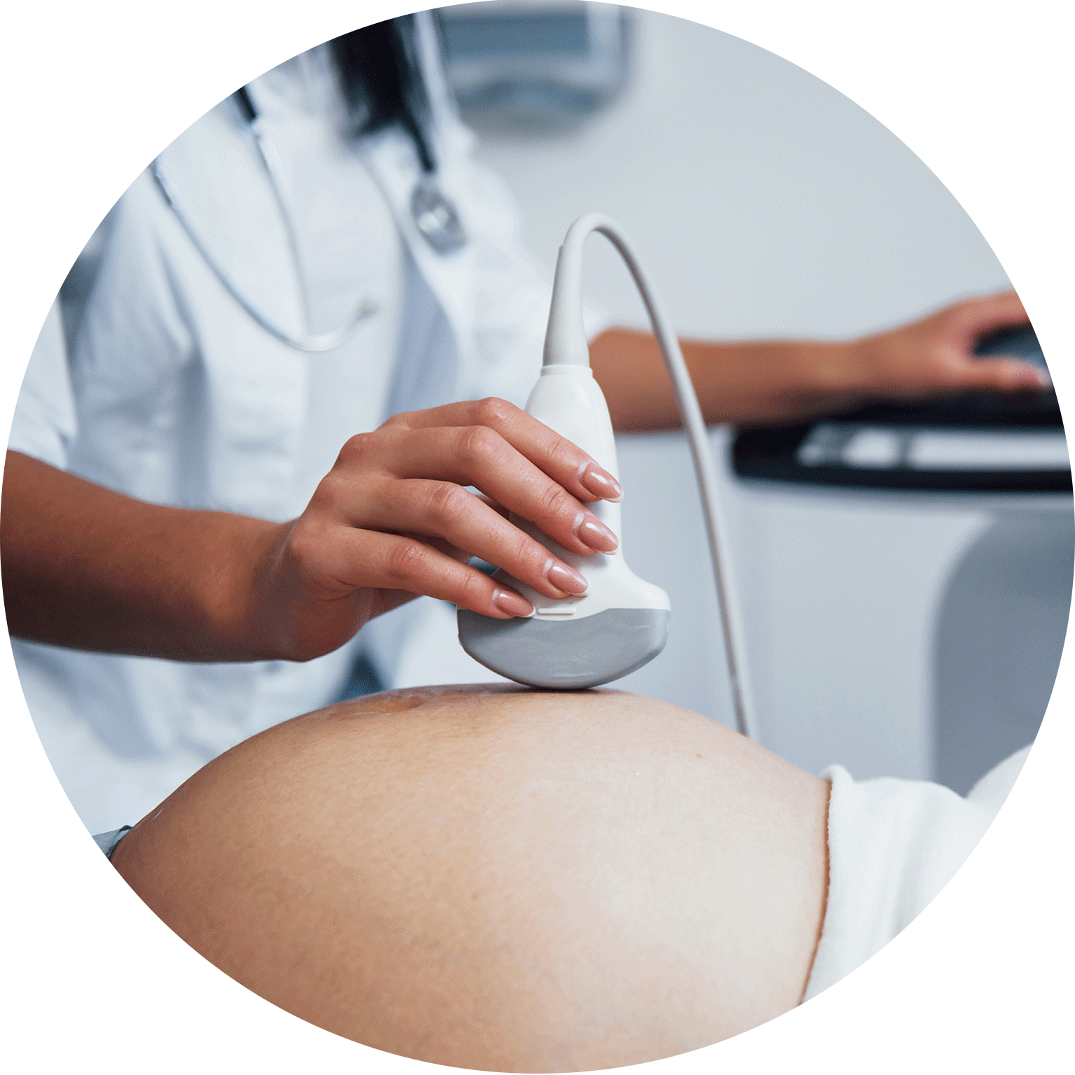
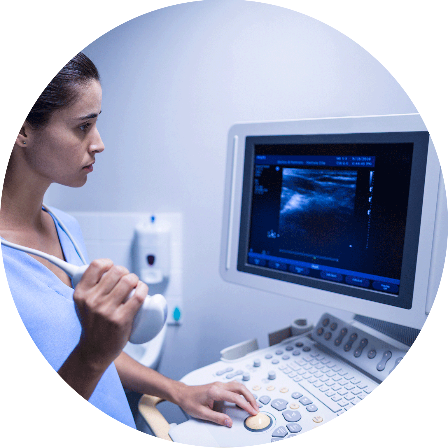
Ultrasound is a medical procedure that uses sound waves to create images of the inside of the body. It is used in the assessment of a variety of conditions, including pregnancy, heart disease, and organ damage. The ultrasound probe is placed on the skin, and the sound waves bounce off the structures inside the body. The image is then displayed on a monitor, providing information about the size, shape, and location of the structures. Ultrasound is a safe and painless procedure that can be used to diagnose a variety of conditions.

Ultrasound scans are used to help diagnose a variety of conditions, including:

– heart problems
– kidney stones
– gallstones
– liver problems
– pancreatitis
– appendicitis
– testicular problems

Things to consider before taking an Ultrasound Scan: 

Before the scan, you will likely be asked to drink plenty of water so that your bladder is full. This is because a full bladder helps to produce clearer images. You may also be asked to avoid eating or drinking for a few hours before the ultrasound so that your stomach and intestines are empty. This is because ultrasound images can be obscured by gas or stool in the intestines. During the ultrasound, you will lie on a bed and a gel will be applied to your skin. A small device called a transducer will be moved over your body, sending out sound waves that create pictures on a screen. The whole process usually takes less than 30 minutes. There is no radiation involved in ultrasound, so it’s generally considered safe for both adults and children.

USG (or) Ultrasound Scan Price at Aarthi Scans and Labs

We have USG with colour doppler scan in the following centres – Chennai (VadapalaniAlwarpetAnna Nagar, Perambur, Tambaram, Tondiarpet, Chrompet, Velachery, Kilpauk, Porur), Tamilnadu (Tirunelveli, Palayamkottai, Madurai, Tenkasi, Tuticorin, Tanjore, Rajapalayam, Kovilpatti), Bangalore (Jayanagar, Indira Nagar).

Note – Scan charges are usually less in towns and cities other than Chennai, and Bangalore.

  • Ultrasound Abdomen and Pelvis, KUB – Rs 900 to 1200
  • Ultrasound Obstetric/Antenatal scan – Rs 900 to 1500 (During I, III trimester)
  • Ultrasound Anomalies scan – Rs 1400 to 2500 (During II trimester)
  • Colour doppler scan (arteries and veins of limbs) – Rs 1500
  • Renal / Carotid / Obstetric doppler – Rs 2500
  • Sonomammogram – Rs 1800 to 2100

WHY SHOULD YOU TAKE AN ULTRASOUND SCAN?

To view the uterus and ovaries during pregnancy &  monitor the developing baby’s health.
To find various other issues like genital and prostrate problems, joint inflammation, gallbladder disease etc.

HOW TO BOOK AN APPOINTMENT IN asl_branchesayappakkamArraypage2

1.

Leave your name & phone number with us

2.

We will reach out to you tounderstand your requirements.

3.

Lab details & charges will beshared with you over the call.

4.

We’ll book your test as per yourpreferred date and time slot.

WHY CHOOSE AARTHI SCANS

35+ Years of Trust

Trusted-by-Doctors-Hospitals

Trusted by Doctors & Hospitals

Honest-Transparent

Honest & Transparent Pricing

Present-in-10-States-and-200-branches

Present in 10+ States and 200+ Branches

1.5-Crore-PATIENTS-SERVED

1.5 CRORE PATIENTS SERVED

FREQUENTLY ASKED QUESTIONS

  • No, ultrasound is not only for pregnancy. It’s a versatile imaging tool used to visualize various organs and tissues in the body, such as the abdomen, pelvis, thyroid, heart, and blood vessels.

  • Yes, ultrasound can detect kidney diseases. It’s commonly used to evaluate the size, shape, and condition of the kidneys and detect abnormalities like kidney stones, cysts, tumors, or structural issues.

  • No, ultrasound is a painless procedure. A gel is applied to the skin, and a transducer is gently moved over the area being examined to create images.

  • Ultrasound works by emitting high-frequency sound waves that bounce off internal structures in the body. These sound waves create echoes, which are then converted into real-time images by a computer.

  • Yes, ultrasound can detect abnormal masses, cysts, or tumors in various organs and tissues. It’s commonly used for breast, thyroid, liver, ovarian, and testicular imaging to identify any abnormal growths.

  • Having a full bladder helps to provide better visualization of pelvic organs, such as the uterus, ovaries, and bladder, during the ultrasound.

  • Transvaginal ultrasound may cause mild discomfort for some women, but it’s generally well-tolerated.

OPG and CBCT In asl_branchesayappakkamArraypage2

mammogram In asl_branchesayappakkamArraypage2

MRI Scan In asl_branchesayappakkamArraypage2

CT Scan In asl_branchesayappakkamArraypage2

Ultrasound In asl_branchesayappakkamArraypage2

Digital X-Ray In asl_branchesayappakkamArraypage2

PET Scan In asl_branchesayappakkamArraypage2

PFT Tests In asl_branchesayappakkamArraypage2

ECG Test In asl_branchesayappakkamArraypage2

DEXA Scan In asl_branchesayappakkamArraypage2

Robotic Lab for Blood tests In asl_branchesayappakkamArraypage2