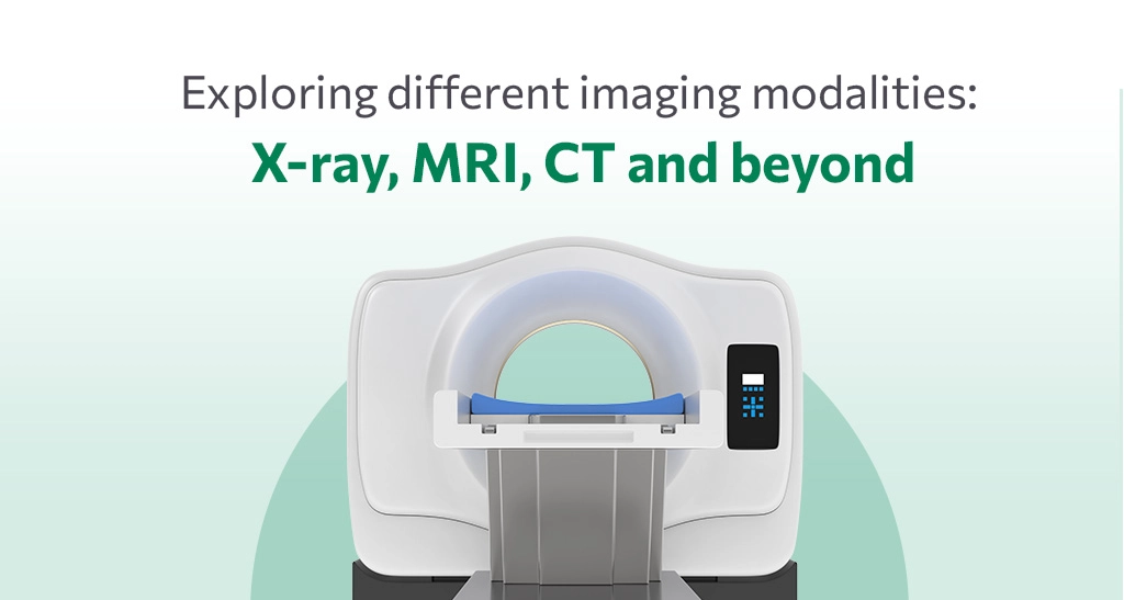
Medical Imaging is the set of procedures done to visualize the internal structures of the body. Medical Imaging is the central part of modern medicine. Imaging modalities are often classified based on the technique used to generate images. The most popular imaging modalities are X-ray, computed tomography (CT) and Magnetic Resonance Imaging (MRI). The development of imaging modalities has been a significant breakthrough in the medical field. Medical Imaging also allows non-invasive detection and treatment of many medical conditions.
| X-ray | X-rays are commonly used imaging tests that produce images of bones and internal organs to reveal signs of diseases and injury. |
| Computed tomography (CT) | Computed tomography (CT) is a non-invasive imaging procedure that also uses X-rays to produce 3-dimensional images of the structures of the body. |
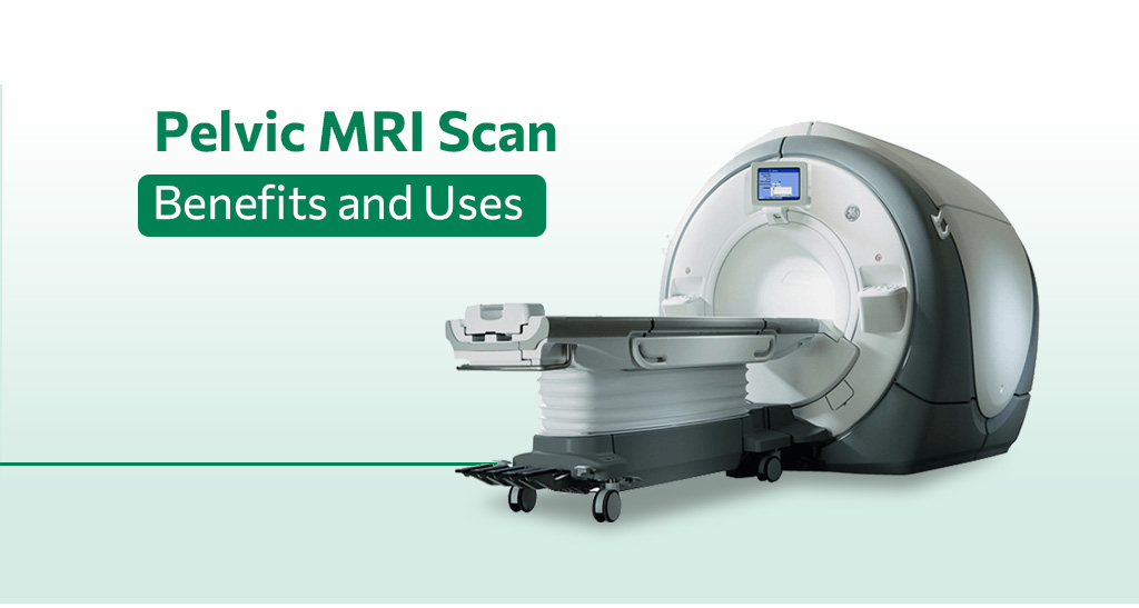
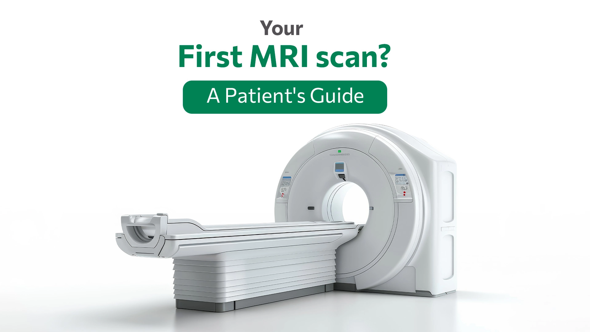
| MRI | MRI or Magnetic Resonance Imaging is an imaging modality used in radiology to form images of the anatomy and physiological processes of the body. that uses a large magnet, radio waves and computer technology to generate clear and detailed images of the body’s organs and structures. |
| Ultrasound | Ultrasound uses sound waves to produce images of structures in the body. Therefore, these images can provide valuable information for diagnosing and assisting treatment for a range of diseases and conditions. |
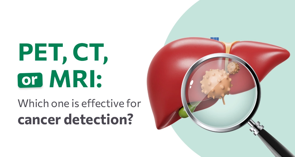
| PET Scan | Positron Emission Tomography (PET) is a medical imaging technique that uses radioactive tracers to visualize the functioning of organs in real-time. PET scans help in managing cancer treatment. |
Common Functions of Imaging Modalities
MRI is a primary diagnostic tool for evaluating skeletal metastases and soft tissue. It diagnoses multiple sclerosis, CNS tumours, brain and spine infections, stroke, injuries in ligaments and tendons, muscle degradation, bone tumours, and blood vessel blockages. Besides anatomy, MRI can also capture physiological processes in the body (functional MRI – fMRI)
CT Scan is predominantly used to diagnose tumours and investigate internal bleeding and injuries. They are particularly useful in identifying acute stroke in the head. Additionally, it helps in the diagnosis, and investigation of the type of stroke, guides surgical interventions and manages the disease.
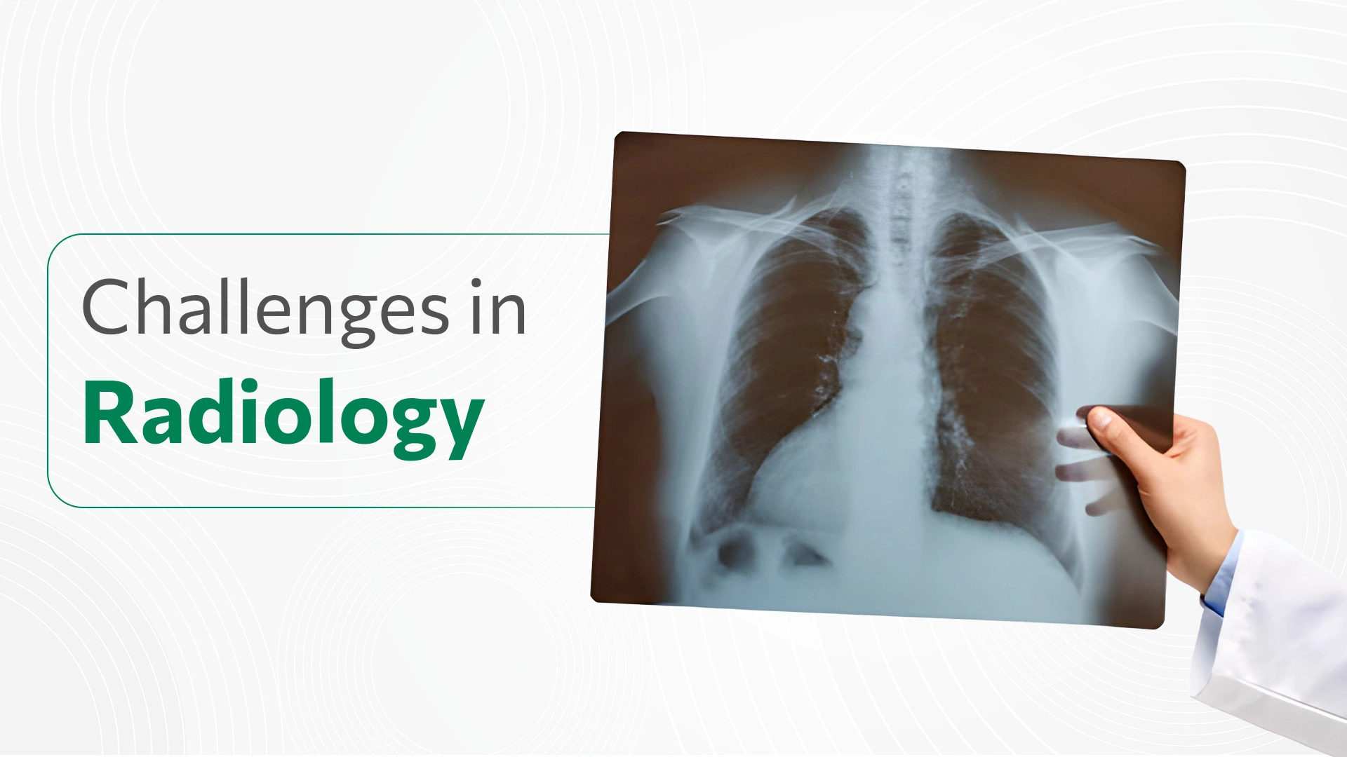
A standard X-ray detects bone fractures, specific injuries, calcifications, and foreign objects, and it is widely used in dentistry.
Mammography, which also utilizes an X-ray beam, produces high-resolution images of the breast, which aids in the detection and monitoring of breast cancer.
PET Scan is often used by oncologists to evaluate cancer and guide cancer treatment. It diagnoses neurosurgical procedures such as tumours, and nervous system disorders, as well as cardiovascular treatment like bypass surgery. It also measures vital functions such as blood flow, blood sugar metabolism etc.
Diagnostic Ultrasound examines various organs within the body such as the abdomen, kidney, pelvis, thyroid, breast etc. Doctors perform Ultrasound when patients experience pain, swelling, or other symptoms that require an internal view of their organs.
Advancements in Medical Imaging
Digital technology has transformed the field of medical imaging. Various medical modalities now offer 3-dimensional images that provide detailed insights into the structures and workings of the body. With this continuous progress in cross-sectional imaging, coupled with the rapid growth & development of interventional radiology has a dramatic impact on patient care & treatment.
- High-resolution CT produces detailed images using very thin slices which helps in many conditions such as lung cancer.
- Computed Tomographic Angiography (CTA) is an X-ray image of blood vessels. It uses an injection of contrast material into your blood vessels and helps diagnose and evaluate blood vessel diseases such as aneurysms or blockages.
- PET/CT evaluates conditions such as epilepsy, Alzheimer’s disease and coronary artery disease.
- Wide bore MRI introduced by Siemens has an 80cm (about 2.62 ft) opening which helps patients with claustrophobia and anxiety of MRIs.
- Cinema Vision MRIs are equipped with a video player where the patient can play a movie/video while undergoing an MRI.
- An early diagnosis of Alzheimer’s disease is possible with PET technology with 93% accuracy. Huntington’s disease is also detectable by a PET scan
In conclusion, medical imaging modalities have revolutionized diagnostics by enabling detailed visualization of internal organs. As technology continues to evolve, imaging modalities are developing, further expanding our ability to diagnose and monitor various medical conditions. The imaging modalities have managed to enhance patient care, improve treatment outcomes and contribute to medical advancements and research significantly.